
Introducing NeuroVista: Truway Health’s Research Platform for Imaging and Connected Medical Data
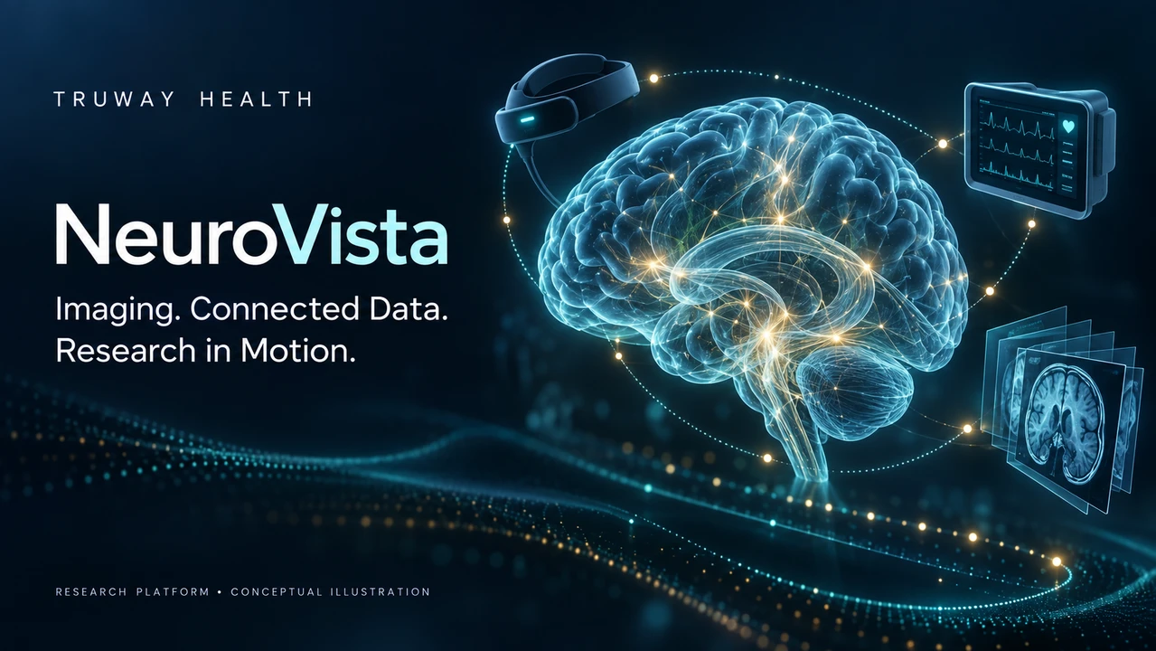
Understanding medical data requires more than a clear image or a single measurement. Researchers also need to understand how information was generated, when it was captured, and which limitations affect its interpretation.
Truway Health is developing NeuroVista, a research software prototype that brings together three-dimensional approach geometry, educational tomography, X-ray diffraction analysis, computer-assisted image segmentation, and Internet of Medical Things—or IoMT—data workflows.
The goal is to create an inspectable environment where researchers can explore computational methods, test assumptions, and identify what needs further validation.
Exploring Imaging and Three-Dimensional Geometry
NeuroVista’s approach-geometry module calculates a straight-line path between an entry point and a target in three-dimensional coordinates. Researchers can examine path length and proximity to point-sampled structures using synthetic examples.
This provides a foundation for studying geometric relationships. It does not account for the full complexity of living anatomy, tissue deformation, instrument dimensions, or intraoperative brain shift. Its current browser visualization is a two-dimensional projection of the geometry, rather than a complete clinical volume viewer.
The tomography module demonstrates how projection data can be reconstructed into an image through filtered back projection. It offers an educational setting for examining reconstruction filters and their effects on synthetic data.
Keeping Materials Analysis Distinct From Patient Imaging
NeuroVista also includes an X-ray diffraction module for research spectra. It detects peaks and estimates crystal-plane spacing using Bragg’s law.
X-ray diffraction serves a different purpose from computed tomography. Within NeuroVista, it supports exploration of materials data; it does not produce patient images or independently identify a material’s composition.
Keeping these functions clearly distinguished helps researchers interpret each output within the method’s actual scope.
A Transparent Starting Point for AI Assistance
NeuroVista’s current image-segmentation module uses normalization and thresholding to generate a proposed mask. It is an inspectable computational baseline, not a trained diagnostic AI model.
That transparency allows researchers to examine how input characteristics and threshold choices change the result. The software returns a review-required flag, although this flag is advisory and does not enforce an approval workflow.
Future development may introduce trained-model adapters, together with documented preprocessing, model versions, evaluation datasets, and uncertainty assessment. Those capabilities would require their own testing before any performance claims could be made.
Extending NeuroVista With Connected-Device Research
The latest build adds an IoMT research module for registering measurement channels and receiving timestamped observations.
Its capabilities include:
- Device registration and channel-specific ingestion credentials.
- Validation of measurement units, values, timestamps, and quality flags.
- Persistent observation storage and duplicate-message handling.
- Dashboard indicators for recent, stale, and poor-quality data.
- Configurable research thresholds and alert acknowledgment.
- A synthetic telemetry simulator for development and testing.
Supported input categories include heart rate, oxygen saturation, temperature, and intracranial pressure. These categories describe the software’s data contract; physical medical-device connections have not yet been integrated or validated.
The module receives observations and does not send treatment commands to devices.
Why Timing Matters
A measurement can arrive now and still describe an earlier state. Network interruptions, delayed transmissions, and clock differences can all complicate a data timeline.
NeuroVista records both observation time and receipt time. It also selects the latest sample by observation time, helping prevent a delayed older reading from replacing a newer one in the dashboard.
These capabilities support Truway Health’s research interest in clinical time and temporal dynamics. Controlled experiments can examine delayed arrivals, missing intervals, duplicate transmissions, and clock offsets to determine how reliably a system represents the sequence of events.
A timestamp alone does not guarantee accurate timing. Clock synchronization and source-device behavior must also be characterized.
What Has Been Tested—and What Comes Next
The v0.2.0 development build passed 16 automated tests across the original computational modules and the IoMT extension, along with a live synthetic-telemetry check.
These results verify selected software behaviors. They do not establish clinical accuracy, patient benefit, hardware compatibility, or readiness for clinical deployment. Browser visual verification also remains outstanding.
Next steps include vendor-specific device adapters, stronger user-access controls, temporal benchmarking, and independent evaluation of data integrity and computational performance.
NeuroVista is a research-use-only prototype. It is not validated for diagnosis, surgical navigation, clinical alarm monitoring, treatment decisions, or direct patient care.
Through NeuroVista, Truway Health is building a practical foundation for exploring how imaging, computation, connected measurements, and time can be studied together—with documented methods and claims that follow the evidence.
Truway Health News & Insights
The Guidewire Is Not a Commodity: The Hidden Safety Architecture of Coronary Care
How precision engineering, clinical intelligence, and medical-device traceability are redefining exp...

Truth Social Suspends @truwayhealth Without Explanation: Truway Health Challenges Digital Censorship and Demands Accountability
October 9, 2026 | Truway Health News & Insights NEW YORK, NY — Truway Health, Inc. is formally chall...

The Stethoscope Standard: Rethinking the Value of U.S. M1 Through Medical Productive Capacity
Money ultimately derives much of its usefulness from confidence: confidence that a dollar can be exc...

Simple Steps for Seniors to Embrace Healthy and Joyful Living
For seniors seeking holistic wellness, healthy aging can feel harder than it should: the body change...

Healthcare Procurement Readiness: Building a More Resilient Clinical Supply Chain
Healthcare supply chains are often judged by a simple question: Did the product arrive? But for hosp...
Pass-Through Chambers and Material Flow in Controlled Healthcare Environments
Controlled healthcare environments depend on more than clean surfaces and filtered air. The way mate...