
Beyond the Lead Apron: Modern Shielding Strategies in Digital Dental Radiography
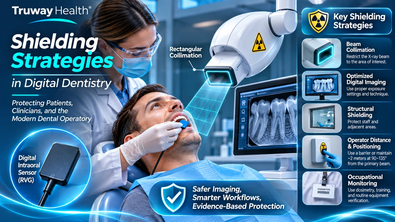
The Evolution of Radiation Protection
For decades, the lead apron became one of the most recognizable symbols of radiation safety in dentistry.
Today, advances in digital radiography and improved radiation-protection science are changing that traditional approach.
Modern radiation protection is increasingly concerned with preventing unnecessary exposure before it occurs, rather than relying primarily on shielding placed over the patient.
The transition is especially relevant to intraoral digital X-ray sensors, which enable rapid image acquisition using electronic detectors.
However, digital technology alone does not guarantee lower exposure. Safety requires the appropriate combination of equipment performance, beam control, positioning, and clinical judgment.
Why Traditional Patient Shielding Recommendations Have Changed
In 2024, the American Dental Association recommended discontinuing routine patient lead aprons and thyroid collars during dental radiographic examinations.
The National Council on Radiation Protection and Measurements reinforced this approach in its 2026 Commentary No. 35 on patient shielding.
The scientific rationale is significant.
Lead aprons do not meaningfully reduce radiation originating from internal scattering within the patient's body. Improperly positioned shielding can also obstruct important anatomy, creating imaging artifacts or requiring repeat exposures.
Instead, greater attention should be directed toward limiting the primary X-ray beam, choosing appropriate examination techniques, and preventing unnecessary images.
This distinction does not eliminate occupational radiation protection. Structural barriers and other protective measures remain important for dental personnel.
Facilities must also comply with applicable radiation-control regulations, which may differ between jurisdictions.
Rectangular Collimation: Controlling the Beam at Its Source
One of the most effective strategies in intraoral radiography is rectangular collimation.
A collimator restricts the dimensions of the X-ray beam before it reaches the patient.
When appropriately implemented, rectangular collimation can substantially reduce the irradiated area compared with traditional circular collimation.
Smaller exposed areas generally mean less unnecessary radiation exposure.
Successful implementation requires careful alignment between the X-ray source and intraoral receptor. Positioning devices can support reproducible geometry and minimize retakes.
Structural Shielding: Protecting the Dental Operatory
Radiation protection must also extend beyond the patient to dental personnel and people occupying neighboring spaces.
A properly designed dental operatory may incorporate protective structural materials, shielded viewing windows, and suitable equipment positioning.
Shielding requirements depend on the X-ray system, anticipated examination workload, room geometry, direction of the radiation beam, and occupancy of adjacent areas.
Different systems may require different protective arrangements.
For this reason, wall composition and barrier design should be evaluated by a qualified radiation-protection professional according to local regulatory requirements.
Digital Sensors and Exposure Optimization
Digital intraoral imaging sensors provide an additional opportunity to improve radiographic workflows.
Their electronic detection capabilities can support efficient acquisition without traditional film processing.
However, proper exposure settings remain essential.
An underexposed digital image may contain excessive noise, while unnecessary exposure increases radiation dose without providing proportional clinical benefit.
Appropriate image quality should therefore be achieved using the lowest exposure consistent with the diagnostic task.
Sensor calibration, correct placement, equipment quality assurance, and operator training are critical components of this strategy.
Handheld Dental X-Ray Systems
Portable and handheld dental X-ray generators present distinct occupational safety considerations.
When permitted by applicable regulations, operators should follow device-specific instructions regarding positioning and protective backscatter shields.
A manufacturer's protective shielding ring must be correctly installed and positioned when required.
Operators should never assume that a portable system is safe to use without its specified protective components.
Building a Modern Radiation Safety Program
Effective radiation protection is not accomplished through one piece of equipment.
It requires an integrated approach combining clinically justified examinations, optimized imaging protocols, verified equipment performance, suitable structural shielding, occupational exposure controls, and staff education.
Quality assurance should include periodic equipment assessments, monitoring where required, and documented corrective actions when performance problems are identified.
As dental imaging systems become more technologically sophisticated, radiation safety programs must continue to evolve alongside them.
The Truway Health Perspective
At Truway Health, the advancement of digital dentistry reinforces the importance of evaluating medical equipment as part of a complete clinical system.
An intraoral sensor cannot be assessed solely by pixel resolution, acquisition speed, or software features.
Its performance must be considered alongside the X-ray generator, beam collimation, imaging technique, patient positioning, regulatory status, and the broader radiation safety environment.
Responsible procurement combines engineering considerations with practical clinical requirements.
The future of radiation protection is not simply about adding more shielding. It is about engineering safer imaging workflows from the beginning.
Explore Medical Imaging Technology
Discover medical technology and dental imaging solutions at TruwayHealth.com.
Educational information only. Radiation-protection systems, shielding construction, and equipment installation should be evaluated according to applicable regulations and qualified professional assessments.
References
-
NCRP Commentary No. 35 — Patient Shielding in Medical Imaging (2026)
-
FDA — Dental Radiographic Examination and Radiation Exposure
Truway Health News & Insights
Beyond the Lead Apron: Modern Shielding Strategies in Digital Dental Radiography
The Evolution of Radiation Protection For decades, the lead apron became one of the most recognizabl...
Digital Dentistry's Quiet Revolution: How Intraoral Sensors Are Transforming Imaging
The Smallest Devices Are Often Behind the Biggest Changes Not every revolution in healthcare arrives...

When Devices Begin to Think: The Next Era of Intelligent Medical Equipment
The Machine Is No Longer Just Measuring. It Is Interpreting. For generations, medical equipment perf...

Medical Supply Case-Pack Economics: How Bulk Purchasing Reduces Healthcare Procurement Costs
Understanding the Economics Behind Medical Supply Purchasing In today's healthcare environment, cont...

The 72-Hour Warning Window: What Biomarkers Can Tell Us About Clinical Deterioration
Introduction: Can the Human Body Signal an Emergency Before It Happens? What if healthcare professio...
Healthcare Equipment Interoperability: Understanding DICOM, HL7, and FHIR
Truway Health | Medical Technology, Digital Infrastructure & Healthcare Innovation Modern medical eq...