
Digital Dentistry's Quiet Revolution: How Intraoral Sensors Are Transforming Imaging
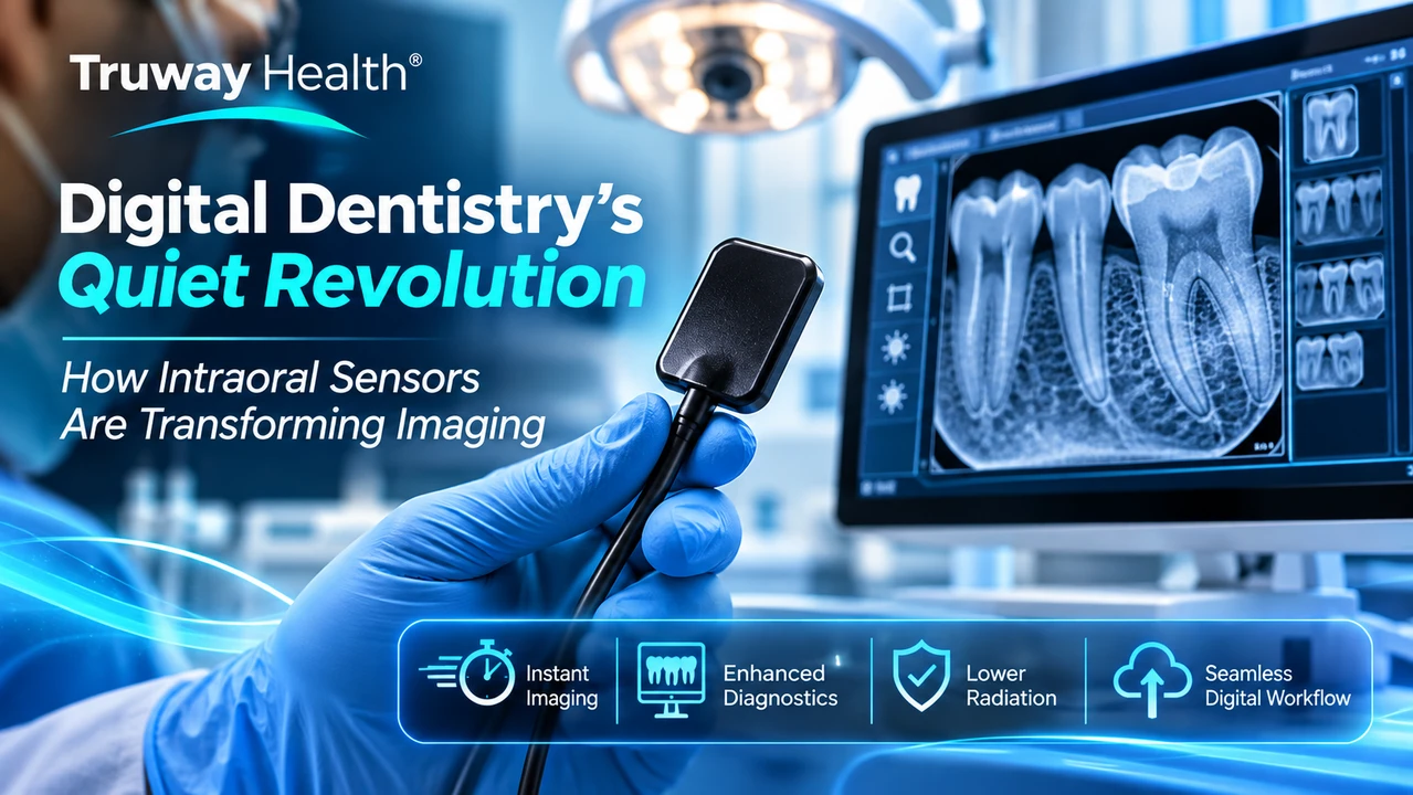
The Smallest Devices Are Often Behind the Biggest Changes
Not every revolution in healthcare arrives as a massive diagnostic machine or a breakthrough surgical system.
Sometimes, it begins with something small enough to fit inside the human mouth.
For decades, dental radiography relied on photosensitive film, chemical processing, and physical image storage. Obtaining a radiograph required multiple steps between X-ray exposure and clinical interpretation.
Today, a compact digital intraoral sensor can acquire radiographic information and deliver an electronic image to a connected workstation within seconds.
This transformation has changed more than the speed of image acquisition.
It has introduced new opportunities for image analysis, electronic recordkeeping, clinical communication, and diagnostic workflow efficiency.
Yet the real technological achievement lies beneath the sensor's exterior: the ability to convert penetrating X-ray photons into digital information suitable for clinical interpretation.
Digital dentistry is not simply replacing film. It is changing what happens to diagnostic information after an image is captured.
1. Understanding the Technology: What Is an RVG Sensor?
RadioVisioGraphy, commonly abbreviated as RVG, refers to a type of digital dental radiography technology.
A digital intraoral sensor functions as an electronic image receptor. It is positioned inside the patient's mouth during a radiographic examination and used with a compatible X-ray generator.
In many solid-state sensors, an X-ray-sensitive scintillator converts incoming X-ray energy into visible light, which is detected by an electronic imaging array.
The resulting electrical signals are processed into a digital image that can be displayed, stored, and reviewed through compatible software.
This process brings together several essential technologies:
-
X-ray detection: Capturing information from radiation transmitted through anatomical structures.
-
Electronic conversion: Translating detected signals into digital image data.
-
Image processing: Adjusting display parameters such as brightness, contrast, and magnification.
-
Computer integration: Transferring images to compatible dental imaging applications.
-
Electronic archiving: Supporting digital storage and retrieval for appropriate clinical documentation.
Importantly, the intraoral sensor itself does not generate X-rays. It detects radiation emitted by a separate, compatible imaging system.
2. CMOS, CCD, and PSP: Three Approaches to Digital Imaging
Digital dental imaging technologies do not all operate in the same manner.
Three established receptor categories are particularly important.
CMOS: Complementary Metal-Oxide-Semiconductor
CMOS sensors employ semiconductor imaging arrays and are widely used in contemporary solid-state dental imaging systems.
Their integration with electronic readout and computer interfaces supports rapid image acquisition.
CCD: Charge-Coupled Device
CCD detectors use a different electronic readout architecture to convert captured information into image data.
CCD technology has played an important historical role in digital dental radiography and remains relevant to understanding the development of modern imaging systems.
PSP: Photostimulable Phosphor Plates
PSP technology uses reusable phosphor plates that store a latent radiographic image.
Unlike solid-state sensors, PSP plates generally require a separate scanning step before the image appears on a workstation.
They can offer a relatively flexible, film-like form factor, although the scanning workflow introduces an additional processing stage.
A 2025 scientific review examined 150 current and historical digital intraoral radiography systems from 55 companies, documenting substantial differences in detector technology, imaging area, resolution, and software characteristics.
The findings reinforce a central lesson for equipment procurement: digital imaging systems are not interchangeable merely because they produce electronic radiographs.
3. Image Resolution: Why More Pixels Do Not Always Mean Better Diagnostics
One of the most frequently advertised characteristics of dental imaging sensors is resolution.
Manufacturers may describe image quality through pixel dimensions, pixel pitch, or spatial resolution measured in line pairs per millimeter (lp/mm).
However, these specifications represent different aspects of imaging performance.
Spatial resolution describes a system's ability to distinguish closely spaced structures.
Pixel pitch represents the physical spacing of image sampling elements.
For idealized digital sampling, theoretical resolution can be approximated as:
Theoretical resolution (lp/mm) = 1 ÷ [2 × pixel pitch (mm)]
For example, a detector with a 0.02 mm pixel pitch has a theoretical sampling limit of 25 lp/mm.
This does not mean the complete imaging system necessarily achieves 25 lp/mm of usable clinical resolution.
Actual performance depends on factors including:
-
Detector construction and electronic noise
-
Scintillator characteristics
-
X-ray focal spot size
-
Imaging geometry and positioning
-
Exposure settings
-
Software processing
-
Display characteristics
Consequently, theoretical resolution should not be confused with measured system performance or proven diagnostic accuracy.
A sophisticated sensor is only one component of a complete dental radiography system.
4. From Image Capture to Clinical Interpretation
Digital intraoral imaging is particularly relevant to several established dental applications.
Bitewing radiography can assist clinicians in assessing interproximal dental caries and evaluating certain changes in supporting alveolar bone.
Periapical radiography provides views of teeth and surrounding structures, supporting assessments involving root anatomy, endodontic treatment, and selected periapical conditions.
Periodontal evaluation may incorporate radiographic findings alongside probing measurements, clinical examination, and patient history.
Restorative and follow-up imaging can help document selected treatment-related findings and support comparisons when examinations are clinically indicated.
Digital systems also allow clinicians to enlarge portions of an image or adjust display parameters for review.
These functions may improve visualization, but they do not create anatomical information that was never captured.
Excessive image enhancement can introduce misleading artifacts.
For this reason, image processing must remain a tool for clinical interpretation rather than a substitute for diagnostic judgment.
5. A New Standard for Responsible Radiation Use
One of the most important developments in dental imaging is the growing emphasis on appropriate examination selection and radiation-dose optimization.
In January 2026, the American Dental Association and the American Academy of Oral and Maxillofacial Radiology published updated recommendations addressing the clinical selection of two-dimensional and three-dimensional dental imaging.
Their central message is straightforward: radiographs should be obtained when clinically justified, rather than automatically performed according to a fixed schedule.
Digital receptors can support lower exposure requirements compared with some conventional film systems, but lower radiation dose is not guaranteed simply because a detector is digital.
Appropriate dose optimization also requires:
-
Clinically justified imaging
-
Suitable exposure parameters
-
Correct sensor positioning
-
Appropriate beam collimation
-
Equipment quality assurance
-
Proper operator training
Digital imaging technology should therefore be evaluated as part of a comprehensive radiation-safety program.
A better image is valuable. An unnecessary exposure is not.
6. The Connected Dental Practice: Why Interoperability Matters
A digital radiograph has little long-term value if it becomes trapped within an isolated software system.
As dental practices adopt electronic records and increasingly connected equipment, compatibility and image portability become important procurement considerations.
The Digital Imaging and Communications in Medicine (DICOM) standard provides a framework for representing and exchanging medical imaging information.
DICOM-related capabilities may assist with imaging interoperability, but actual integration depends on the specific equipment, software implementation, and supporting systems.
In March 2026, the American Dental Association emphasized the need to improve interoperability between dental imaging platforms and electronic records.
For dental practices, this raises several practical questions.
Can images be exported without loss of clinically relevant information?
Can another authorized provider access them through an appropriate exchange process?
Can a practice migrate its imaging archive when changing software vendors?
Will future equipment remain compatible with the existing digital environment?
USB connectivity alone does not establish full software interoperability, and the ability to export a common image format does not necessarily indicate complete DICOM compliance.
The future of digital dentistry will depend as much on the movement of information as on the technology used to capture it.
7. The Often-Overlooked Engineering Challenges
Although intraoral sensors are compact, they operate in a demanding clinical environment.
Repeated positioning, cable movement, cleaning, barrier placement, and routine handling all impose requirements on device design.
Important engineering considerations include mechanical durability, electrical safety, liquid-ingress resistance, connector integrity, sensor dimensions, and serviceability.
Patient comfort is another major factor.
Sensor thickness, active imaging area, rounded edges, and positioning accessories can influence ease of use and the ability to capture clinically useful images.
Infection prevention is equally important.
The Centers for Disease Control and Prevention classifies digital intraoral radiography sensors as semicritical devices.
CDC guidance calls for appropriate FDA-cleared barrier protection and reprocessing according to device capabilities and manufacturer instructions, with additional cleaning and intermediate-level disinfection requirements for sensors that cannot tolerate sterilization or high-level disinfection.
A disposable barrier is not, by itself, a complete infection-control program.
These operational considerations illustrate why purchasing decisions should extend well beyond advertised image resolution.
8. The Economics of Digital Dentistry: Looking Beyond Purchase Price
For dental practices, acquiring a digital imaging system is an investment in a long-term clinical workflow.
A useful financial comparison should consider the full lifecycle of ownership.
That includes the initial equipment cost, imaging software licenses, computer compatibility, technical support, sensor replacement, protection accessories, training, and expected downtime.
Digital imaging may eliminate film-development chemicals and reduce certain processing tasks.
However, electronic equipment introduces expenses associated with software management, hardware maintenance, cybersecurity, and eventual replacement.
A sensor that appears economical at purchase may become costly if replacement components are unavailable or the system cannot integrate with existing practice software.
For this reason, procurement teams should evaluate the total operating environment rather than comparing equipment prices in isolation.
9. Artificial Intelligence: The Next Layer of Dental Imaging
The next stage of digital dental imaging may increasingly involve computational assistance with image interpretation.
AI-enabled systems are being developed and evaluated for applications such as identifying radiographic features associated with dental caries, periodontal bone changes, and other conditions.
Such tools may provide a second analytical perspective or help organize information for clinical review.
But automated findings are not equivalent to independently confirmed diagnoses.
Algorithm performance can vary according to image quality, training data, patient characteristics, and clinical setting.
The ADA's 2025 technical report on artificial intelligence in dental image analysis emphasizes external validation, data quality, patient privacy, and preservation of clinical judgment.
The most promising future is not necessarily automated dentistry.
It is dentistry in which reliable imaging and carefully validated computational tools support qualified clinicians in making better-informed decisions.
10. The Truway Health Perspective: Building Better Imaging Workflows
At Truway Health, digital dental imaging represents an important intersection of medical equipment engineering, clinical practice, and healthcare technology procurement.
The value of an intraoral sensor extends beyond the ability to produce an electronic X-ray image.
Successful clinical deployment requires compatibility with an appropriate X-ray generator, reliable image-acquisition software, validated performance, suitable infection-control procedures, and ongoing equipment support.
Healthcare organizations should also examine documentation, model-specific regulatory status, installation requirements, and software integration before acquiring equipment for clinical use.
Truway Health's medical equipment catalog includes digital dental imaging technologies, reflecting the broader movement toward electronically connected diagnostic workflows.
As technology evolves, responsible equipment selection will remain essential.
The goal is not simply to make dental imaging faster. It is to make the entire imaging process more reliable, accessible, interoperable, and clinically useful.
Conclusion: The Revolution Is in the Information
The transition from film to digital intraoral radiography may appear modest compared with other advances in medical technology.
Yet its effects reach far beyond the dental operatory.
Compact sensors have helped transform image acquisition, enabled electronic diagnostic workflows, and created new opportunities for information sharing and computational analysis.
The next generation of dental imaging will likely place greater emphasis on interoperability, equipment durability, image quality, and carefully validated AI assistance.
These developments must remain grounded in patient safety, clinical evidence, and responsible radiation use.
The quiet revolution in dentistry is not about a single device.
It is about transforming a moment of radiation exposure into dependable diagnostic information that can be interpreted, protected, and used responsibly throughout the patient's care.
Explore Digital Dental Imaging at Truway Health
Explore Truway Health's Greatlife HD Digital RVG Intraoral X-Ray Sensor and other healthcare technologies available through TruwayHealth.com.
Product-specific specifications, software compatibility, regulatory documentation, and clinical-use requirements should be verified before deployment.
Truway Health, Inc. — Medical Technology, Clinical Research & Healthcare Supply Solutions
Disclaimer: This article is intended for educational and industry information. It does not replace professional dental judgment, clinical practice guidelines, or applicable equipment regulatory requirements.
Scientific and Regulatory References
-
American Dental Association — 2026 Dental Imaging Patient Selection Recommendations
-
FDA — Selection of Patients for Dental Radiographic Examinations
-
Intraoral Digital Radiography: Comprehensive Review of Technical Specifications (2025)
Truway Health News & Insights
Beyond the Lead Apron: Modern Shielding Strategies in Digital Dental Radiography
The Evolution of Radiation Protection For decades, the lead apron became one of the most recognizabl...
Digital Dentistry's Quiet Revolution: How Intraoral Sensors Are Transforming Imaging
The Smallest Devices Are Often Behind the Biggest Changes Not every revolution in healthcare arrives...

When Devices Begin to Think: The Next Era of Intelligent Medical Equipment
The Machine Is No Longer Just Measuring. It Is Interpreting. For generations, medical equipment perf...

Medical Supply Case-Pack Economics: How Bulk Purchasing Reduces Healthcare Procurement Costs
Understanding the Economics Behind Medical Supply Purchasing In today's healthcare environment, cont...

The 72-Hour Warning Window: What Biomarkers Can Tell Us About Clinical Deterioration
Introduction: Can the Human Body Signal an Emergency Before It Happens? What if healthcare professio...
Healthcare Equipment Interoperability: Understanding DICOM, HL7, and FHIR
Truway Health | Medical Technology, Digital Infrastructure & Healthcare Innovation Modern medical eq...