
3D Ultrasound vs. Traditional Ultrasound: What Is the Difference?
Introduction
Ultrasound is one of the most widely used medical imaging technologies. It uses high-frequency sound waves to create images of structures inside the body without using ionizing radiation.
While traditional 2D ultrasound remains an important diagnostic tool, advances in imaging technology have introduced 3D ultrasound, which can capture and reconstruct volumetric information. These technologies are closely related, but they are not interchangeable in every clinical situation.
So, what exactly is the difference between 3D and traditional ultrasound—and when might one approach provide information that the other cannot?
What Is Traditional 2D Ultrasound?
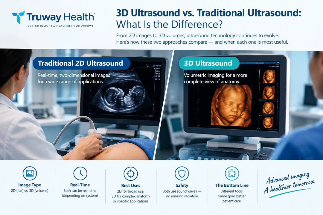
Traditional ultrasound generally produces a two-dimensional, real-time image of the body.
During an examination, an ultrasound transducer sends sound waves into the body. The returning echoes are processed by the ultrasound system and displayed as an image.
Depending on the examination, ultrasound can be used to evaluate structures such as:
- The heart
- Blood vessels
- Abdominal organs
- Thyroid and other superficial structures
- Muscles and tendons
- Reproductive organs
- Soft tissues
- Fetal anatomy during pregnancy
Traditional ultrasound is particularly valuable because it can provide real-time imaging while a clinician moves the transducer across the area being examined.
What Is 3D Ultrasound?
3D ultrasound expands on conventional ultrasound by acquiring multiple image planes and combining them into a three-dimensional volume.
Instead of representing anatomy as a single flat image, the system can construct a volumetric representation that can be manipulated or reviewed from different perspectives.
Depending on the equipment and examination, 3D ultrasound may allow users to:
- Acquire volumetric datasets
- Review anatomy in multiple planes
- Rotate reconstructed images
- Examine spatial relationships between structures
- Store volumetric information for later review
- Generate surface or multiplanar renderings
Some systems can also incorporate 4D ultrasound, which essentially refers to 3D imaging displayed dynamically over time.
3D vs. 2D Ultrasound at a Glance
| Feature | Traditional 2D Ultrasound | 3D Ultrasound |
|---|---|---|
| Image representation | Two-dimensional | Three-dimensional volume |
| Real-time imaging | Yes | Depending on system |
| Spatial information | Primarily a selected imaging plane | Volumetric |
| Multiple-plane review | Limited during acquisition | Possible from acquired volume |
| Data storage | Individual images/clips | Volumetric datasets |
| Equipment complexity | Generally lower | Generally higher |
| Operator skill | Important | Important, particularly for volume acquisition |
| Common applications | Broad diagnostic use | Specialized imaging and volumetric assessment |
| Ionizing radiation | None | None |
| Clinical usefulness | Established across many applications | Application-dependent |
How Does 3D Ultrasound Work?
The fundamental physics are similar.
An ultrasound transducer sends sound waves into the body and receives returning echoes. The system processes those echoes to estimate the location and characteristics of tissue interfaces.
The key difference is how the imaging information is acquired and reconstructed.
A conventional examination may acquire an image plane such as:
X–Y
A volumetric acquisition adds another spatial dimension:
X–Y–Z
The resulting dataset can potentially be viewed from different orientations.
This can be especially useful when anatomy has a complex three-dimensional structure.
When Can 3D Ultrasound Be Useful?
The value of 3D imaging depends heavily on the clinical question.
1. Obstetric and gynecologic imaging
3D ultrasound can provide volumetric information about reproductive anatomy and fetal structures.
In appropriate circumstances, the additional spatial information may help clinicians visualize anatomy from planes that are difficult to obtain directly with conventional 2D scanning.
Importantly, 3D ultrasound does not automatically provide a better diagnostic examination than 2D ultrasound. The appropriate technique depends on the clinical indication and the expertise of the imaging professional.
2. Cardiac imaging
Three-dimensional echocardiographic techniques can provide volumetric information about cardiac structures.
This can potentially assist with assessment of:
- Cardiac chambers
- Valve anatomy
- Structural abnormalities
- Cardiac volume
- Spatial relationships between cardiac structures
However, conventional 2D echocardiography remains an important component of many cardiac examinations.
3. Musculoskeletal imaging
Volumetric imaging may help characterize certain structures involving:
- Tendons
- Muscles
- Ligaments
- Joints
- Soft-tissue abnormalities
The ability to review a volume from multiple perspectives can sometimes complement conventional imaging.
4. Procedural and interventional applications
Ultrasound is increasingly used to guide procedures because clinicians can visualize anatomy in real time.
Depending on the equipment and procedure, 3D or volumetric imaging may provide additional spatial information.
What About Color Doppler?
Color Doppler is different from 3D imaging.
This distinction is important.
3D ultrasound describes the spatial representation of anatomy.
Color Doppler provides information related to moving blood, using Doppler principles to estimate characteristics of blood flow.
A modern ultrasound system may combine several capabilities, including:
2D imaging + Color Doppler + 3D imaging
These are complementary technologies rather than competing versions of the same feature.
For example, a clinician might use conventional grayscale imaging to examine anatomy and then use Doppler imaging to evaluate blood flow.
Is 3D Ultrasound Better Than 2D Ultrasound?
Not necessarily.
The better question is:
Which imaging method is most appropriate for the clinical question?
Traditional 2D ultrasound has several important advantages. It is widely available, versatile, relatively fast, and capable of producing excellent real-time images.
3D ultrasound adds volumetric information, but acquiring and interpreting a useful 3D dataset can require appropriate equipment, technique, and clinical expertise.
A three-dimensional image can also look impressive without necessarily providing additional diagnostic information.
Therefore, more dimensions do not automatically mean better diagnosis.
Advantages of 3D Ultrasound
Potential advantages include:
Volumetric information
Instead of relying exclusively on a single imaging plane, clinicians can potentially evaluate a stored volume.
Multiplanar visualization
A volume can potentially be reconstructed into different viewing planes.
Improved spatial understanding
Complex anatomy may be easier to understand when its three-dimensional relationships can be explored.
Dataset storage
A volumetric dataset can potentially be preserved for subsequent review, depending on the system and workflow.
Educational applications
Three-dimensional representations can be useful for teaching anatomy and explaining certain findings.
Limitations of 3D Ultrasound
3D imaging also has limitations.
Image quality depends on acquisition
A poor-quality acquisition cannot necessarily be rescued by reconstruction.
Motion can affect datasets
Patient movement, breathing, cardiac motion, or fetal movement can influence volumetric acquisitions depending on the application.
Operator expertise remains important
Advanced technology does not eliminate the importance of appropriate scanning technique.
Not every clinical question requires 3D
Many examinations can be effectively performed using conventional 2D ultrasound.
Equipment and workflow
3D-capable systems may involve additional hardware, software, training, or processing requirements.
Is Ultrasound Safe?
Ultrasound does not use X-rays or other forms of ionizing radiation.
Instead, diagnostic ultrasound uses mechanical sound waves.
Medical ultrasound is generally considered an established diagnostic imaging modality when appropriately used. As with any medical technology, however, ultrasound should be performed for an appropriate clinical purpose and according to applicable safety principles.
Patients should discuss specific imaging decisions with their healthcare professional.
The Future of Ultrasound: More Than Just Better Images
The evolution of ultrasound is not simply about moving from 2D to 3D.
Modern systems increasingly combine:
- High-resolution imaging
- Color Doppler
- Spectral Doppler
- 3D and volumetric imaging
- Automated measurements
- Artificial intelligence-assisted analysis
- Cloud and network connectivity
- DICOM interoperability
- Portable and point-of-care systems
This creates an opportunity to think about ultrasound as a multimodal clinical information platform, rather than simply an imaging device.
The next generation of ultrasound technology may increasingly focus on how imaging data can be integrated with other clinical information while maintaining appropriate privacy, security, validation, and clinical oversight.
Choosing the Right Ultrasound Technology
For healthcare organizations evaluating an ultrasound platform, specifications should be considered in the context of intended use.
Important questions include:
- What anatomical structures will be examined?
- Is real-time 2D imaging sufficient?
- Is Color Doppler required?
- Is volumetric/3D imaging clinically necessary?
- Which transducers are needed?
- Does the system support the desired workflow?
- Can images be exported using appropriate standards such as DICOM?
- What quality-control and calibration procedures are available?
- What training is required?
- Has the system been appropriately evaluated for its intended clinical use?
The transducer configuration can be just as important as the imaging software. Convex, linear, phased-array, endocavitary, and other probes serve different applications.
The Bottom Line
Traditional 2D ultrasound and 3D ultrasound are complementary technologies.
2D ultrasound provides highly useful real-time imaging across a broad range of clinical applications.
3D ultrasound adds volumetric information that can allow anatomy to be explored from multiple perspectives and may be particularly useful for selected applications.
Neither technology should be considered universally superior. The appropriate approach depends on the clinical question, anatomy, equipment, acquisition technique, operator expertise, and intended use.
As ultrasound continues to evolve, the most meaningful advances may come not simply from adding another imaging dimension, but from combining high-quality imaging with Doppler, quantitative analysis, interoperability, artificial intelligence, and clinically validated workflows.
About Truway Health
At Truway Health, we are interested in the intersection of medical technology, diagnostic imaging, digital health, and clinical research. Understanding the capabilities—and limitations—of emerging technologies is an important part of responsible healthcare innovation.
Medical technology should ultimately serve the clinical question, the healthcare professional, and the patient.
This article is for educational purposes and is not medical advice. Ultrasound examinations and interpretation should be performed by appropriately trained healthcare professionals, and clinical decisions should be based on the complete clinical context.
Truway Health News & Insights

Moving from RN to Family Nurse Practitioner What You Need to Know
Experienced registered nurses often reach a point where strong bedside skills still leave a lingerin...

The Guidewire Is Not a Commodity: The Hidden Safety Architecture of Coronary Care
How precision engineering, clinical intelligence, and medical-device traceability are redefining exp...

Truth Social Suspends @truwayhealth Without Explanation: Truway Health Challenges Digital Censorship and Demands Accountability
October 9, 2026 | Truway Health News & Insights NEW YORK, NY — Truway Health, Inc. is formally chall...

The Stethoscope Standard: Rethinking the Value of U.S. M1 Through Medical Productive Capacity
Money ultimately derives much of its usefulness from confidence: confidence that a dollar can be exc...

Simple Steps for Seniors to Embrace Healthy and Joyful Living
For seniors seeking holistic wellness, healthy aging can feel harder than it should: the body change...

Healthcare Procurement Readiness: Building a More Resilient Clinical Supply Chain
Healthcare supply chains are often judged by a simple question: Did the product arrive? But for hosp...