
Theranostics: Combining Diagnostic Imaging and Targeted Treatment
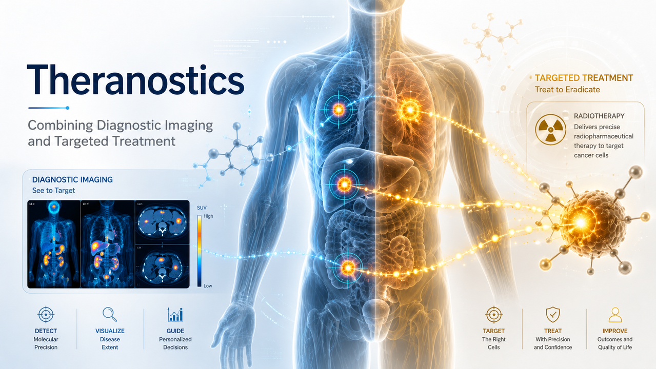
Modern medicine is moving beyond treating every patient with the same standardized approach. One of the most promising examples is theranostics, a precision-medicine strategy that combines diagnostic testing with targeted treatment.
The term blends two words: therapeutics and diagnostics. In nuclear medicine, theranostics typically uses two closely related radiopharmaceuticals. One is designed to locate and image a specific biological target, while the other delivers radiation directly to cells expressing that same target.
This “find it, confirm it, and treat it” model may help clinicians select appropriate patients, identify the location and extent of disease, deliver treatment more precisely, and monitor how the disease responds.
What Is a Radiopharmaceutical?
A radiopharmaceutical is a medicine containing a radioactive isotope attached to a molecule that travels to a particular tissue, receptor, protein, or cellular process.
The targeting molecule may be a:
- Small molecule
- Peptide
- Antibody
- Protein
- Nanoparticle
- Receptor-binding ligand
For diagnostic imaging, the radiopharmaceutical emits radiation that can be detected by a PET or SPECT camera. The resulting images show where the agent accumulates inside the body.
For treatment, a similar targeting molecule carries a therapeutic radionuclide. Instead of primarily producing an image, it releases radiation intended to damage or destroy the targeted cells.
Targeted radionuclide therapy is a form of systemic radiation treatment because the medicine travels through the bloodstream. Unlike external-beam radiation, it can potentially reach multiple disease sites throughout the body during the same treatment.
How Theranostics Works
A typical theranostic pathway includes several connected steps.
1. Identify a molecular target
Clinicians first identify a receptor, protein, transporter, or metabolic pathway that is strongly expressed by the disease.
An effective target should ideally be present on the abnormal cells while having lower or manageable expression in healthy tissue.
2. Perform diagnostic imaging
A small amount of a diagnostic radiopharmaceutical is administered. The agent travels through the body and binds to cells carrying the intended target.
A PET, PET/CT, SPECT, or SPECT/CT scanner detects the radiation emitted by the tracer and produces a map of its distribution.
This imaging may help determine:
- Whether the target is present
- How strongly different lesions express it
- Where the disease has spread
- Whether the patient may be eligible for targeted treatment
- Which normal organs may also absorb the agent
3. Deliver targeted therapy
When the diagnostic study confirms adequate target expression, a therapeutic version of the radiopharmaceutical may be administered.
The targeting component recognizes the same molecular feature identified during imaging, while the therapeutic radioisotope delivers radiation to the targeted tissue.
4. Monitor distribution and response
Follow-up laboratory testing and imaging may be used to evaluate treatment distribution, response, organ function, and possible toxicity.
This connected process distinguishes theranostics from treatment decisions based only on general disease classification.
Current Clinical Applications
Theranostic principles have been used in nuclear medicine for decades. Radioactive iodine is a foundational example because thyroid cells naturally absorb iodine. Diagnostic iodine imaging can identify iodine-avid tissue, while iodine-131 may be used to treat certain thyroid cancers.
More recent applications include neuroendocrine tumors and prostate cancer.
Neuroendocrine tumors
Some gastroenteropancreatic neuroendocrine tumors express somatostatin receptors. Diagnostic PET agents can visualize these receptors and help determine whether receptor-targeted radionuclide treatment may be appropriate.
Lutetium Lu 177 dotatate is FDA-approved for adults and pediatric patients aged 12 years and older with somatostatin receptor-positive gastroenteropancreatic neuroendocrine tumors.
Prostate cancer
Prostate-specific membrane antigen, or PSMA, is expressed at elevated levels in many prostate cancers. PSMA-targeted PET imaging can identify tumors that express the target and support treatment selection.
Lutetium Lu 177 vipivotide tetraxetan delivers radiation to PSMA-expressing cells. In March 2025, the FDA expanded its indication for certain adults with PSMA-positive metastatic castration-resistant prostate cancer following androgen-receptor pathway inhibitor therapy. Patient selection requires an approved PSMA PET imaging product to confirm appropriate tumor expression.
Potential Benefits of Theranostics
More informed patient selection
The diagnostic scan provides direct evidence that the intended target is present before treatment begins. This may reduce the likelihood of using a target-specific therapy when the disease does not adequately express that target.
Whole-body disease mapping
PET and SPECT imaging can reveal target-positive disease throughout the body, including lesions that may not be apparent through symptoms or conventional anatomical imaging alone.
Targeted radiation delivery
The radiopharmaceutical is designed to concentrate radiation in target-expressing tissues. Although normal organs may still receive radiation, the goal is to create a more favorable distribution than would occur with untargeted systemic exposure.
Personalized treatment planning
Imaging may provide information about radiopharmaceutical uptake, retention, tumor burden, and normal-organ exposure.
Researchers are increasingly examining patient-specific dosimetry—the calculation of absorbed radiation dose—to determine whether treatment activity and scheduling can be better individualized. FDA guidance also emphasizes dosage optimization during the clinical development of oncology radiopharmaceutical therapies.
Treatment-response monitoring
Follow-up imaging, laboratory measurements, symptoms, and clinical outcomes can be evaluated together to determine whether the disease is responding and whether additional treatment should be considered.
Risks and Limitations
Theranostics is targeted, but it is not completely selective.
Healthy organs that express the same molecular target or participate in eliminating the radiopharmaceutical may also receive radiation. Depending on the agent, organs requiring attention may include the kidneys, liver, bone marrow, salivary glands, lacrimal glands, or gastrointestinal system.
Potential adverse effects vary by radiopharmaceutical and may include:
- Fatigue
- Nausea
- Temporary blood-count reductions
- Dry mouth
- Changes in kidney or liver function
- Bone-marrow suppression
- Secondary radiation-related effects
- Infertility or reproductive risks
- Delayed toxicity
The amount of target expression may also differ among lesions within the same patient. A person may have some tumors that strongly absorb the agent and others that do not. Tumors can also change over time or after previous treatment.
Careful review of diagnostic imaging, blood counts, kidney function, liver function, prior treatment, disease burden, medications, and overall health is therefore necessary before treatment. Current research continues to examine adverse-event management, dosimetry, supply limitations, workforce requirements, reimbursement, and long-term safety.
The Role of PET and SPECT Imaging
PET and SPECT do more than identify the anatomical location of a tumor. They can reveal biological activity based on the selected radiotracer.
PET imaging detects pairs of photons produced following positron emission. It is widely used for high-sensitivity molecular imaging and quantitative measurements such as standardized uptake values.
SPECT imaging detects gamma photons emitted directly from a radiotracer. SPECT may be useful for diagnostic imaging, treatment verification, and post-therapy assessment with certain radionuclides.
CT or MRI may be combined with these techniques to provide anatomical detail. Together, molecular and anatomical imaging can show both where a lesion is located and how it behaves biologically.
Emerging Areas of Research
Theranostics is expanding beyond its established targets. Researchers are investigating radiopharmaceuticals directed at:
- Fibroblast activation protein
- Chemokine receptors
- Integrins
- HER2
- Gastrin-releasing peptide receptors
- Carbonic anhydrase IX
- Immune-system targets
- Tumor-associated enzymes
- Components of the tumor microenvironment
New therapeutic isotopes are also being evaluated. Beta-emitting radionuclides can provide radiation across a measurable tissue range, while alpha emitters release highly energetic particles over shorter distances. Each approach presents different opportunities and safety considerations.
Other developing areas include nanoparticle carriers, antibody-based radiopharmaceuticals, combination immunotherapy, artificial intelligence-assisted image analysis, spatial dosimetry, and computational “digital twin” models intended to simulate individual treatment response.
Theranostics and Precision Medicine
The central value of theranostics is not simply that one product can image and another can treat. Its importance lies in connecting several clinical decisions through the same molecular target.
The diagnostic phase asks:
Can we see the target?
The therapeutic phase asks:
Can we use that target to deliver treatment?
The monitoring phase asks:
Did the treatment reach the disease, and how did the patient respond?
This feedback loop may support more personalized cancer care by linking imaging, biology, radiation delivery, treatment response, and safety monitoring.
Theranostics does not eliminate uncertainty, and it is not suitable for every patient or every cancer. However, it represents an important shift toward treatments selected according to measurable biological characteristics rather than diagnosis alone.
As radiopharmaceutical research advances, theranostics may continue to expand into additional cancers and other diseases. Its long-term success will depend on validated molecular targets, reliable isotope production, standardized imaging, individualized dosimetry, rigorous clinical trials, long-term toxicity monitoring, and access to trained multidisciplinary teams.
Medical disclaimer: This article is provided for general educational purposes and does not constitute medical advice, diagnosis, or a treatment recommendation. Radiopharmaceutical imaging and therapy must be ordered, administered, and monitored by appropriately qualified healthcare professionals. Approved indications, eligibility requirements, risks, and availability vary by product and patient.
Truway Health News & Insights

Moving from RN to Family Nurse Practitioner What You Need to Know
Experienced registered nurses often reach a point where strong bedside skills still leave a lingerin...

The Guidewire Is Not a Commodity: The Hidden Safety Architecture of Coronary Care
How precision engineering, clinical intelligence, and medical-device traceability are redefining exp...

Truth Social Suspends @truwayhealth Without Explanation: Truway Health Challenges Digital Censorship and Demands Accountability
October 9, 2026 | Truway Health News & Insights NEW YORK, NY — Truway Health, Inc. is formally chall...

The Stethoscope Standard: Rethinking the Value of U.S. M1 Through Medical Productive Capacity
Money ultimately derives much of its usefulness from confidence: confidence that a dollar can be exc...

Simple Steps for Seniors to Embrace Healthy and Joyful Living
For seniors seeking holistic wellness, healthy aging can feel harder than it should: the body change...

Healthcare Procurement Readiness: Building a More Resilient Clinical Supply Chain
Healthcare supply chains are often judged by a simple question: Did the product arrive? But for hosp...